C3. All cells have a lipoprotein cell membrane. In eukaryotic cells it is attached to the cytoskeleton.
Student Outcome: C3.1
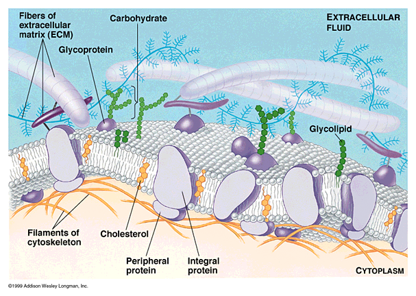
Describe the structure and function of the cell membrane in terms of the fluid mosaic model.
Fluidity of the Plasma Membrane
- At body temperature, the phospholipid bilayer has consistency of olive oil.
- In each single part of the bi-layer, the phospholipid tails "wiggle", and entire phospholipid molecules can move sideways at a rate of about 2 µm — the length of a prokaryotic cell — per second.
- Phospholipid molecules rarely flip-flop from one layer to the other.
- Fluidity of the phospholipid bilayer allows cell membranes to be flexible.
- Some proteins are held in place by cytoskeletal filaments; most drift in fluid bilayer.

The Membrane Is a Mosaic
- Cell membrane and organelle membranes have unique proteins; Red Blood Cell plasma membrane contains 50 or more types of proteins.
- Membrane proteins determine most of the membrane’s functions.
- Channel proteins allow a particular molecule to cross membrane freely (e.g., Cl- channels).
- Carrier proteins selectively interact with a specific molecule so it can cross the plasma membrane (e.g., Na+ - K+ pump, sodium potassium pump).
- Receptor proteins are shaped so a specific molecule (e.g., hormone or other molecule)can bind to it.
- Enzymatic proteins catalyze specific metabolic reactions
Source: www.sirinet.net
Very, very nice video of the fluid mosaic in action - showing diffusion and hormone activation!
![]()
Go here for an amazing video of how viruses infect cells - how they are able to get into and out of infected cells.
Student Outcome: C3.2
Describe the role of the membrane in endocytosis and exocytosis.
If the molecules are too large to pass through the cell membrane, a portion of the cell membrane forms an invagination that contains some of the extracellular fluid and materials. This invagination pinches off to form a new, membrane-enclosed organelle with the cell cytoplasm. The cell can then utilize the materials. This process if called endocytosis. There are three subtypes of endocytosis: phagocytosis (cell eating), pinocytosis (cell drinking) and receptor- mediated endocytosis, which involves interaction of specific molecules in the extracellular fluid with specific membrane protein receptor causing the membrane to invaginate.
Exocytosis, the reverse of endocytosis, serves as a mechanism for the secretion of cellular products in the extracellular environment. Proteins and other molecules produced within the cell are first packaged within the Golgi apparatus. These vesicles are then fused with the cell membrane and release into the extracellular environment. For example, chemical transmitters from nerve endings are released in this manner.
Source: http://science.csustan.edu/flora/zool2232/Transport%20Lab.htm
- Endocystosis, is a general term for the process whereby very large particles of material are wrapped with plasma membrane and moved into the cell in the form of vesicles or vacuoles. None of the trapped material actually moves through the membrane, but remains on the other side of the original membrane, even while the vacuole is inside the cell.
- Exocytosis, is the reverse of endocytosis. Quatities of material are expelled from the cell without ever passing through the membrane as individual molecules. By using the processes of endocytosis and exocytosis, some specialized types of cells move large amounts of bulk material into and out of themselves.

Source: http://www.brooklyn.cuny.edu/bc/ahp/LAD/C5/C5_Endocytosis.html
Great movie on the creation of Golgi body.
http://vcell.ndsu.nodak.edu/animations/proteintrafficking/movie.htm
More movies on endocytosis
![]()
Student Outcome: C3.3
State three functions of the cytoskeleton.
A three-dimensional network of microtubules and filaments that provides internal support for the cells, anchors internal cell structures, and functions in cell movement and division.
Source: http://www.emc.maricopa.edu/faculty/farabee/BIOBK/BioBookglossC.html
CYTOSKELETON
Just as your body depends on your skeleton to maintain its shape and size, so a Cell needs structures to maintain its shape and size. In Animal Cells, an internal framework called CYTOSKELETON maintains the Shape of the Cell. THE CYTOSKELETON MAINTAINS THE THREE-DIMENSIONAL STRUCTURE OF THE CELL, PARTICIPATES IN THE MOVEMENT OF ORGANELLES WITHIN THE CYTOSOL, AND HELPS THE CELL MOVE. The Cytoskeleton is a network of long protein strands located in the Cytosol, that are Not surrounded by a membrane. The CYTOSKELETON consists of Three Types: MICROTUBULES MICROFILAMENTS AND INTERMEDIATE FILAMENTS.

MICROTUBULES
Microtubules are HALLOW TUBES like plumbing pipes. They are the Largest Strands of the Cytoskeleton. Microtubules are made of a PROTEIN called TUBULIN.
Microtubules have THREE FUNCTIONS:
- To maintain the shape of the cell and hold organelles in place.
- To serve as tracks for organelles and molecules to move along within the cell.
- When the Cell is about to divide, two short cylinders of Microtubules at right angles known as Centrioles can be found situated in the cytoplasm near the nuclear envelope. Centrioles organize the micortubules of the cytoskeleton during Cell Division in animal cells, plant cells lack centrioles.
MICROFILAMENTS
MICROFILAMENTS are NOT HALLOW and have a structure that resembles ROPE made of
TWO TWISTED CHAINS OF PROTEIN called ACTIN. MICROFILAMENTS can CONTRACT, causing movement. Muscle Cells have many microfilaments.
This video shows a white blood cell (neutrophil) "chasing" a bacterium - movement made possible by these filaments.
![]()
Here is another video showing how Ameoba move - again only through the action of the microfilaments.
INTERMEDIATE FILAMENTS
Intermediate filaments are rods that anchor the nucleus and some other organelles to their place in the cell. They maintain the internal shape of the nucleus. Hair-follicle (hair-root) cells produce large quantities of intermediate filament proteins. These proteins make up most of the hair shaft - your hair!
Here is a picture of intermediate filaments of a rat kangaroo's epithelial cell as seen through a fluorescence optical microscope.

Source: www.sirinet.net