C2. There are two main types of cell organisation.
Student Outcome: C2.1
Compare the size and structural organisation of prokaryotic and eukaryotic cells.
| Characteristic | Prokaryotes | Eukaryotes |
| Size of cell | Typically 0.2-2.0 m m in diameter | Typically 10-100 m m in diameter |
| Nucleus | No nuclear membrane or nucleolus (nucleoid) | True nucleus, consisting of nuclear membrane & nucleolus |
| Membrane-enclosed organelles | Absent | Present; examples include lysosomes, Golgi complex, endoplasmic reticulum, mitochondria & chloroplasts |
| Flagella | Consist of two protein building blocks | Complex; consist of multiple microtubules |
| Glycocalyx | Present as a capsule or slime layer | Present in some cells that lack a cell wall |
| Cell wall | Usually present; chemically complex (typical bacterial cell wall includes peptidoglycan) | When present, chemically simple |
| Plasma membrane | No carbohydrates and generally lacks sterols | Sterols and carbohydrates that serve as receptors present |
| Cytoplasm | No cytosketeton or cytoplasmic streaming | Cytoskeleton; cytoplasmic streaming |
| Ribosomes | Smaller size (70S) | Larger size (80S); smaller size (70S) in organelles |
| Chromosome (DNA) arrangement | Single circular chromosome; lacks histones | Multiple linear chromosomes with histones |
| Cell division | Binary fission | Mitosis |
| Sexual reproduction | No meiosis; transfer of DNA fragments only (conjugation) | Involves meiosis |
Source: http://www.life.umd.edu/classroom/bsci424/BSCI223WebSiteFiles/ProkaryoticvsEukaryotic.htm
Here is a reasonable video showing some of them main structures of a plant cell.
![]()
This is a famous and very realistic animation of a cell called: Inner life of a Cell.
![]()
Student Outcome: C2.2
Describe the structure and function of the following organelles: nucleus, mitochondrion, chloroplast, vacuole, Golgi body, and endoplasmic reticulum.
- Nucleus
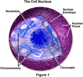
This is where the DNA is kept and RNA is transcribed. RNA is transported out of the nucleus through the nuclear pores. Proteins needed inside the nucleus are transported in through the nuclear pores. The nucleolus is usually visible as a dark spot in the nucleus (note the dark nucleolus in this electron microscope photo of a nucleus), and is the site of ribosome formation.
- Mitochondria

Mitochondria (singular: mitochondrion) are the sites of aerobic respiration, and generally are the major energy production center in eukaryotes. Mitochondria have two membranes, an inner and an outer, clearly visible in this electron microscope photo of a mitochondrion. Note the reticulations, or many infoldings, of the inner membrane, This serves to increase the surface area of membrane on which membrane-bound reactions can take place. The existence of this double membrane has led many biologists to theorize that mitochondria are the descendants of some bacteria that was endocytosed by a larger cell billions of years ago, but not digested. This fascinating theory of symbiosis, which might lend an explanation to the development of eukaryotic cells, has additional supporting evidence. Mitochondria have their own DNA and their own ribosomes; and those ribosomes are more similar to bacterial ribosomes than to eukaryotic ribosomes.
- Chloroplasts

These organelles (item number 6 in the above SEM picture) are the site of photosynthesis in plants and other photosynthesizing organisms. They also have a double membrane. There is a more complete description of the chloroplast here, in the chapter on photosynthesis.
- Golgi apparatus

This organelle modifies molecules and packages them into small membrane bound sacs called vesicles. These sacs can be targetted at various locations in the cell and even to its exterior.
Go here (University of Utah: Learning Genetics site) for some more information about vesicles - some stunning videos are included.
- Endoplasmic Reticulum (ER)
(see above diagram)
The ER is the transport network for molecules targeted for certain modifications and specific final destinations, as opposed to molecules that are destined to float freely in the cytoplasm. There are two types of ER, rough and smooth. Rough ER has ribosomes attached to it, and smooth ER does not.
Source: web.mit.edu
This is from the famous John Kyrk website which has lots of fabulous animations - this one is on the cell. You can click on each organelle to get more detail.
This site shows a comparison between prokaryotic, animal and plant cells - use the magnifying glass to see more details.
This BBC site compares cell structure with the structure of cities.
This Open University site acts like a digital microscope - needs flash and be patient.
Really nice animation of a cell from Learn.Genetics and the University of Utah. You just point at one part of the cell and they will show you an animation for how it work. Click on another button and the cell changes to a plant cell.
Student Outcome: C2.3
Understand why even the simplest cell has several hundred genes.
Scientists Find Smallest Number Of Genes Needed For Organism's Survival
CHAPEL HILL - The minimum number of protein-producing genes a single-celled organism needs to survive and reproduce in the laboratory is somewhere between 265 and 350, according to new research directed by a top University of North Carolina at Chapel Hill scientist.
Using a technique known as global transposon mutagenesis, Dr. Clyde A. Hutchison III, professor of microbiology at the UNC-CH School of Medicine, and colleagues at The Institute for Genomic Research (TIGR) in Rockville, Md., found that roughly a third of the genes in the disease-causing Mycoplasma genitalium were unnecessary for the bacterium's survival.
Such research is a significant step forward in creating minimal, tailor-made life forms that can be further altered for such purposes as making biologically active agents for treating illness, Hutchison said. More immediately, it boosts scientists' basic understanding of the question, "What is life?"
"Surprisingly, the minimal set of genes we found included about 100 whose function we don't yet understand. This finding calls into question the prevailing assumption that the basic molecular mechanisms underlying cellular life are understood, at least broadly."
Source: http://www.sciencedaily.com/releases/1999/12/991213052506.htm